The De Winter ECG pattern has been reported to indicate acute left anterior descending coronary artery occlusion and is often considered to be an ST elevation myocardial infarction STEMI equivalent. The de Winter ECG pattern is a recently-described STEMI equivalent that emergency physicians and paramedics must be aware of.

De Winter Syndrome An Underrecognized Electrocardiography Finding In Myocardial Infarction Journal Of Emergency Medicine
These patients typically have critical stenosis of the LAD requiring emergent PCI or thrombolysis.
. The De Winter ECG pattern has been reported to indicate acute left anterior descending coronary artery occlusion and is often considered to be an ST elevation myocardial infarction STEMI equivalent. These patients were more likely to be younger and male and tended to have hypercholesterolaemia. We present a patient with acute proximal LAD occlusion who presented with an evolutionary ECG change in which the de Winter.
We aimed to investigate the morphology of the De Winter ECG pattern and evaluate the test characteristics of the De Winter pattern for the diagnosis of acute coronary occlusion. The reported positive predictive value PPV for the de Winter ECG pattern to predict an acute left anterior descending artery LAD lesion is inconsistent. This pattern should be treated as being equivalent to an anterior STEMI.
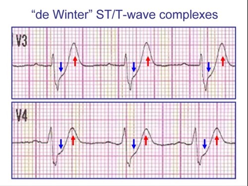
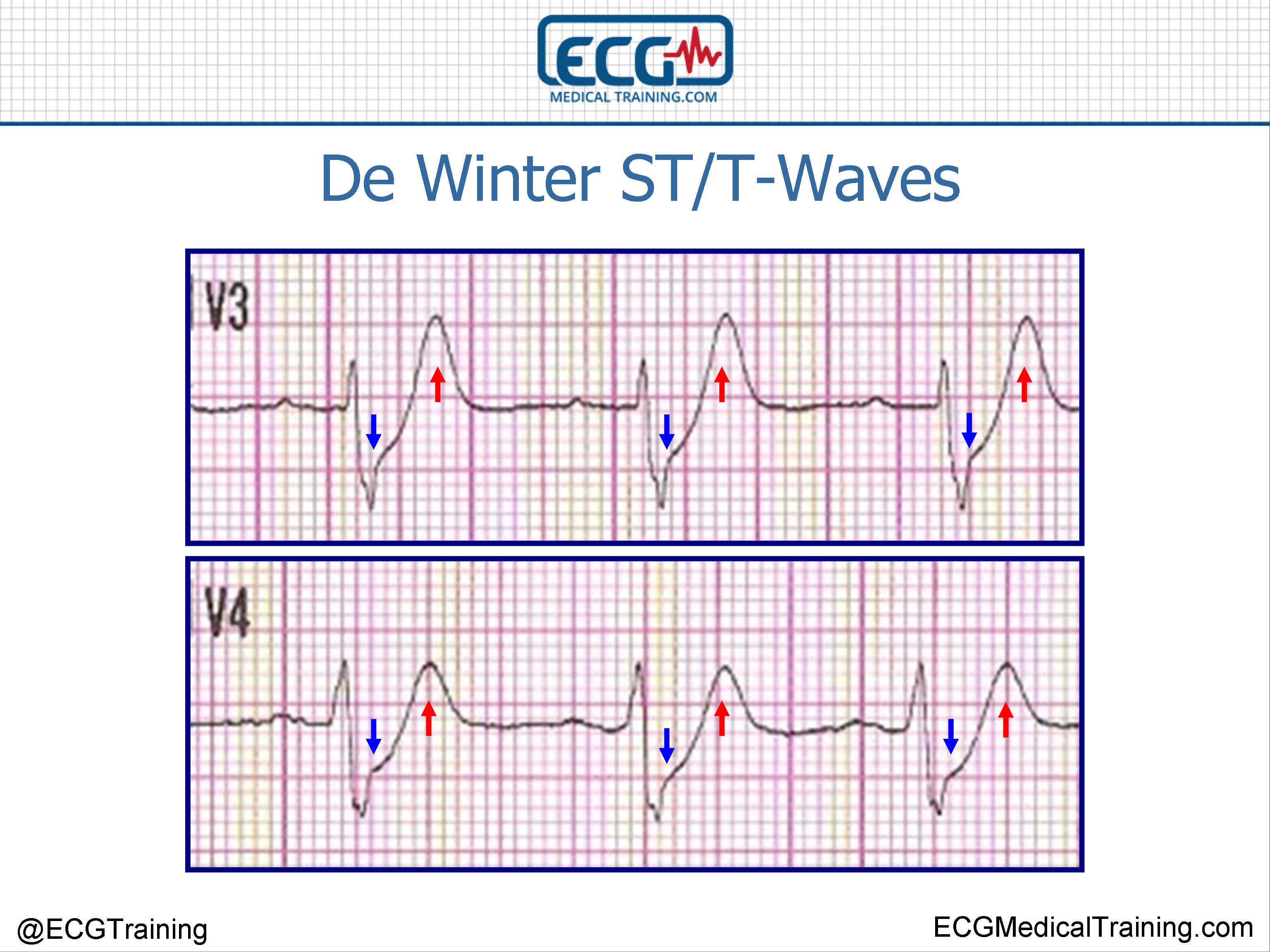
T waves associated with De Winters are often referred to as being. ECG characteristics of De Winters T waves include. Differentiation of this ECG pattern is essential to refer patients for appropriate and immediate reperfusion therapy.
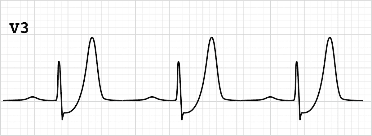
Tall often very prominent T waves in the precordial leads. Besides the morphology of upsloping or nonupsloping ST depression STD may have different significance of severity and prognostication. The De Winter ECG pattern has been reported to indicate acute left anterior descending coronary artery occlusion and is often considered to be an ST elevation myocardial infarction STEMI equivalent.
De Winter in 2008 the de Winter ECG pattern is an anterior STEMI equivalent that presents without obvious ST segment elevation. We aimed to investigate the morphology of the De Winter ECG pattern and evaluate the test characteristics of the De Winter pattern for the diagnosis of acute coronary occlusion. The distinct ECG pattern was identified in approximately 2 of patients with LAD stenosis in that study also.
In 2008 de Winter et al described an ECG pattern suggesting that it should be considered an ST-elevation myocardial infarction STEMI equivalent de Winter Verouden Wellens Wilde 2008 with the potential to predict critical stenosis or occlusion of the left anterior descending coronary artery LAD. We report a case of a 34yearold man with a history of smoking who presented. Hence hyperacute T-waves are the first ECG change in STE-ACSSTEMI.
We aimed to investigate the morphology of the De Winter ECG pattern and evaluate the test characteristics of the De Winter pattern for the diagnosis of acute coronary occlusion. Recall that T-waves should not exceed 10 mm in chest leads and 5 mm in limb leads. De Winter Rd et al A new sign of proximal lad occlusion NEJM 2008359.
Very slight 05-1mm ST elevation in lead aVR. This de Winter ECG pattern is not static but can evolve from hyperacute T waves. Since they are short-lived it is uncommon to encounter them in clinical practice.
The main characteristics of the de Winter electrocardiogram ECG pattern are up-sloping ST-segment depression in the V 1 to V 6 leads followed by tall and symmetrical T waves which remain consistent with no evolutionary ECG changes. De Winters sign persistent hyperacute T-wave syndrome As mentioned above hyperacute T-waves have a short duration. The de Winter electrocardiography ECG pattern is a sign that implies proximal left anterior descending coronary artery occlusion in patients with chest pain.
The de Winter ECG pattern indicates occlusion of the LAD and is frequently underrecognized by clinicians which increases morbidity and mortality. These patients are suffering occlusion myocardial infarction OMI and require immediate reperfusion therapy. First reported by Dutch Professor of Cardiology Robbert J.
The de Winter electrocardiogram pattern is a transient electrocardiographic phenomenon that presents at early stage of ST-segment elevation myocardial infarction Clin Cardiol 41 2018 pp. More than 1mm of ST depression in the precordial leads.
De Winter St T Waves Ecg Medical Training
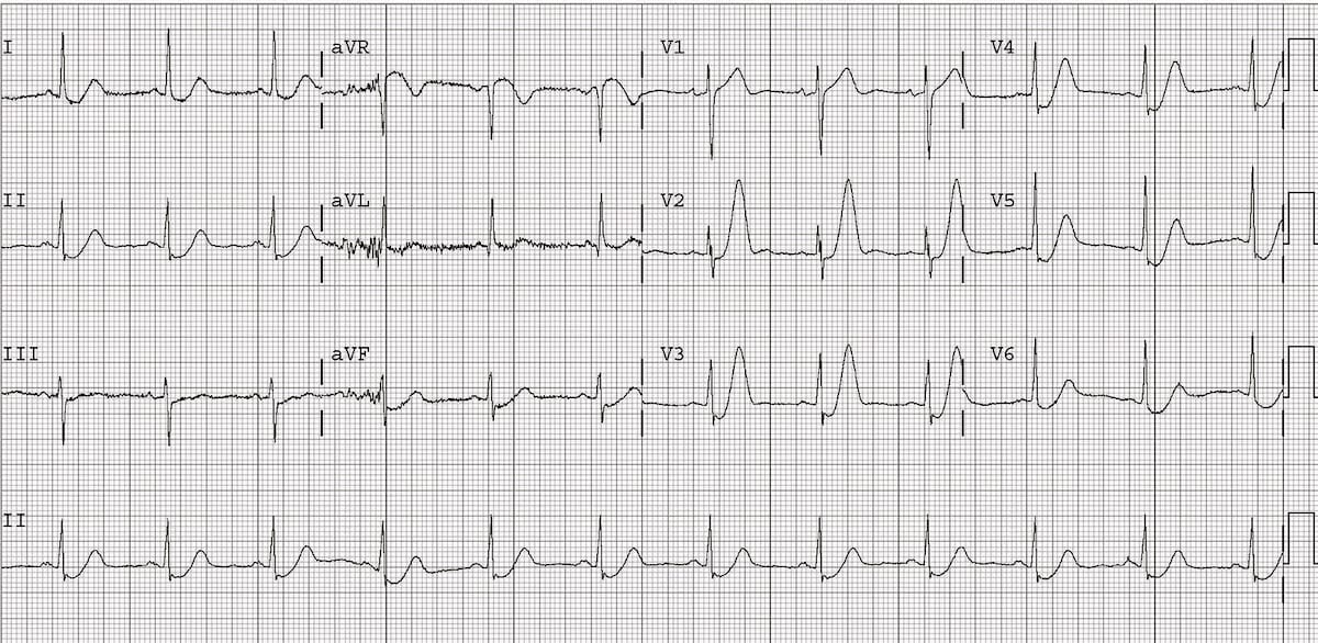
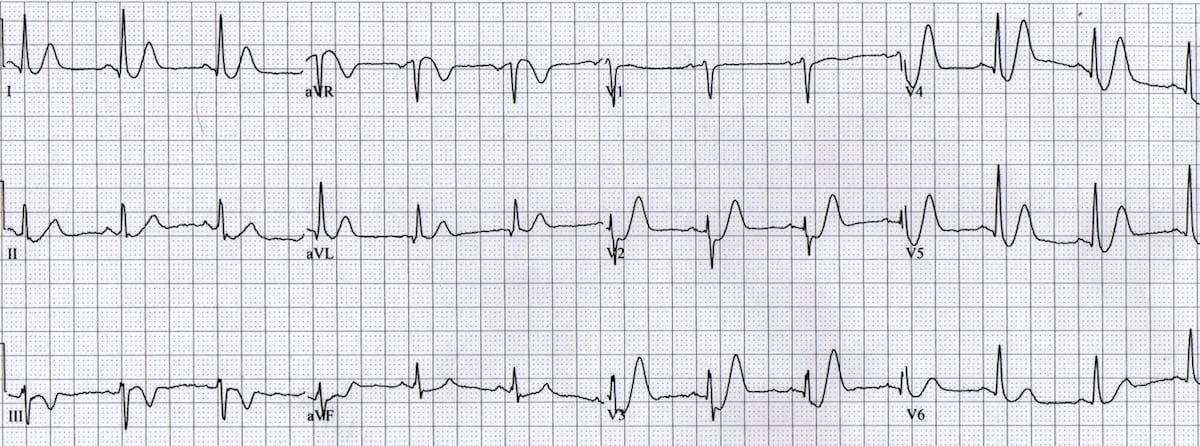
De Winter T Wave Litfl Ecg Library Diagnosis
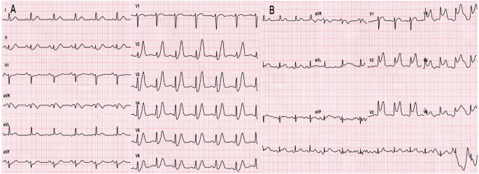
Evolutionary De Winter Pattern From De Winter Ecg To Stemi A Case Report Bmc Cardiovascular Disorders Full Text
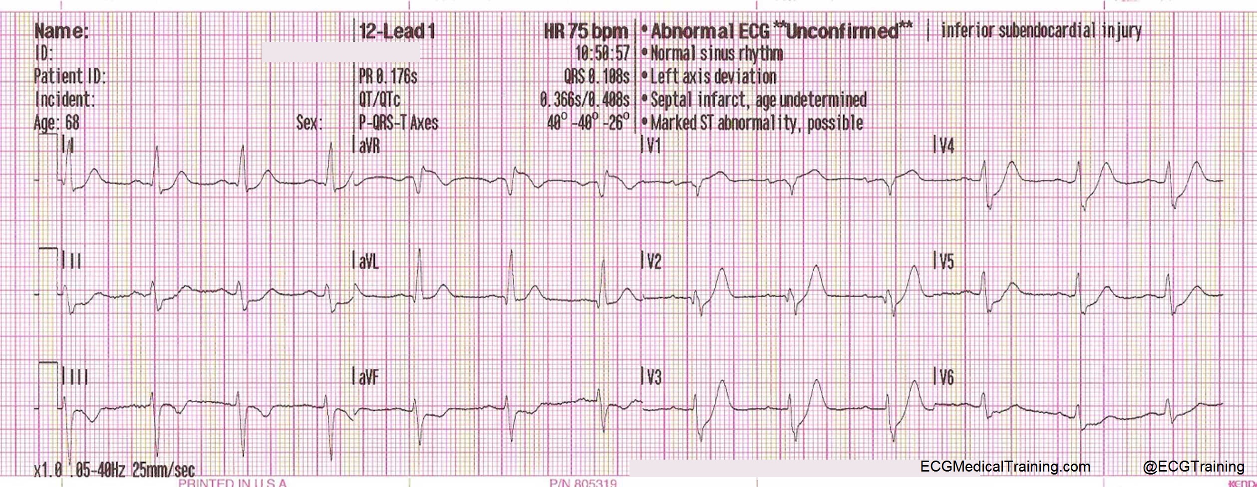
De Winter St T Waves Ecg Medical Training

De Winter T Wave Litfl Ecg Library Diagnosis
De Winter T Wave Litfl Ecg Library Diagnosis
De Winter T Wave Litfl Ecg Library Diagnosis
Dewinter S T Waves
0 comments
Post a Comment